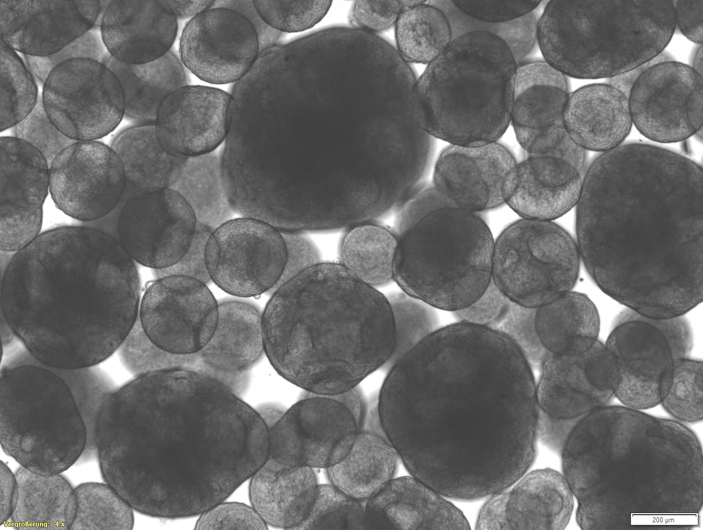
We establish reporter spheroids to detect the activation of cellular signaling pathways. These are suited, for example, to be used in microfluidic systems for the sensor-controlled real-time analysis of in-vitro cell and tissue cultures.
Reporter Spheroids

Our development: reporter spheroids for analyzing cell reactions in real time

Our reporter spheroids are generated from customized reporter cells. If a specific signaling pathway is activated in these cells, this can be read out quickly and easily in real time via the formation of a reporter protein that has been stably integrated into the genome of the cells using a reporter construct.
If these reporter spheroids are linked to other cell and tissue models via microfluidic platforms such as organ-on-a-chip systems, cell stress-inducing and toxic substance effects in particular can be analyzed in real time.
Our reporter spheroids can be used to quantify inflammatory markers such as cytokines or the activation of sensitizing signaling pathways in upstream in-vitro tissue and cell cultures, especially patient material, in a highly sensitive manner, even in the course of time.

Privacy warning
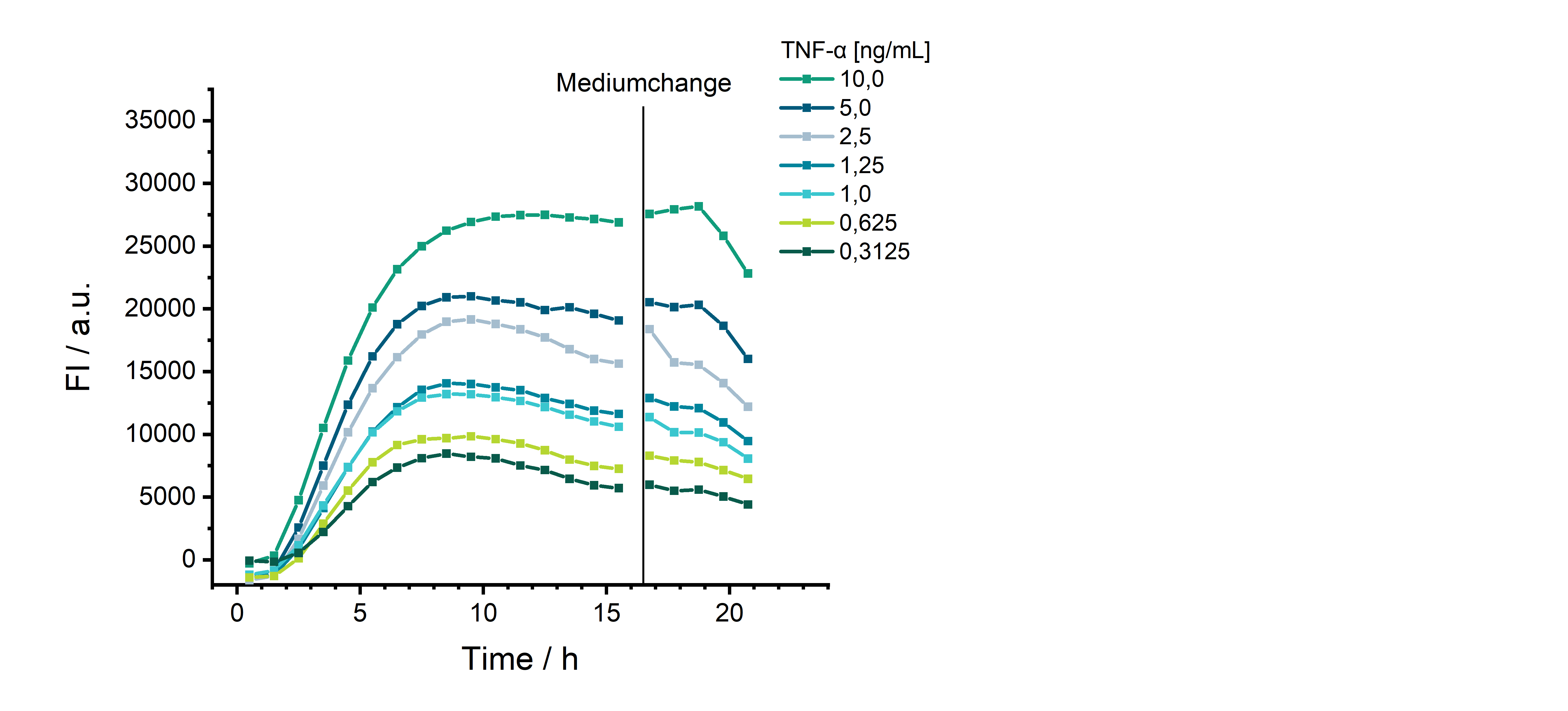
With the click on the play button an external video from www.youtube.com is loaded and started. Your data is possible transferred and stored to third party. Do not start the video if you disagree. Find more about the youtube privacy statement under the following link: https://policies.google.com/privacyStimulation of the reporter spheroids with TNF-α leads to a concentration-dependent increase in fluorescence intensity over time. This makes these reporter spheroids suitable for real-time detection of the inflammation marker TNF-α in the culture medium of tissue models under investigation. Images courtesy of Fraunhofer IZI-BB and Fraunhofer IGB

Applications
We provide a broad portfolio of spheroids as in-vitro test systems for various applications in drug development and pre-clinical testing as well as in disease modeling, especially in cancer research.
With our spheroids, reporter spheroids and tumoroids, we address various applications for
- Assessment of the efficacy and safety of drugs and chemicals (development and preclinical testing),
- Determination of toxicity,
- Analysis of transport processes and absorption of a substance,
- Investigation of the activation of cellular signaling pathways (sensitization, inflammation) and
- Disease research, especially cancer research
Tabbed contents
Portfolio
Our portfolio of spheroids
We provide specifically suitable 3D microtissues for various issues and applications:
- Spheroids based on different cell lines
- Reporter spheroids for non-invasive real-time detection of the activation of cellular signaling pathways
- Tumoroids based on different tumor cell lines
Services
Range of services
We offer the following services relating to our spheroids, which we carry out in state-of-the-art laboratories on behalf of and in collaboration with customers:
- Conducting studies and substance screenings
- Development of innovative (reporter) spheroids including
- Production of genetically modified cell lines (stable and transient)
- Reporter cell lines with different intracellular and secreted reporters
- Cultivation of spheroids from (tumor) cell lines
- Phenotypic and functional characterization of models
Analysis methods
Analysis methods
In our 3D microtissues, the effects of chemicals, active substances and products on the architecture of the tissue and physiological properties can be investigated. We offer the following analysis methods:
- Vitality measurements, for example using the MTT test (colorimetric test), alamarBlue(TM)
- Multiplex analysis for the detection and quantification of secreted proteins such as cytokines, chemokines, growth factors and antimicrobial peptides
- Histological and immunohistochemical staining of tissue thin sections
- Protein analyses using Western blot
- Analysis of specific signaling pathway activation via reporter proteins
- RNA and DNA analyses using PCR or qPCR and sequencing
 Fraunhofer Institute for Interfacial Engineering and Biotechnology IGB