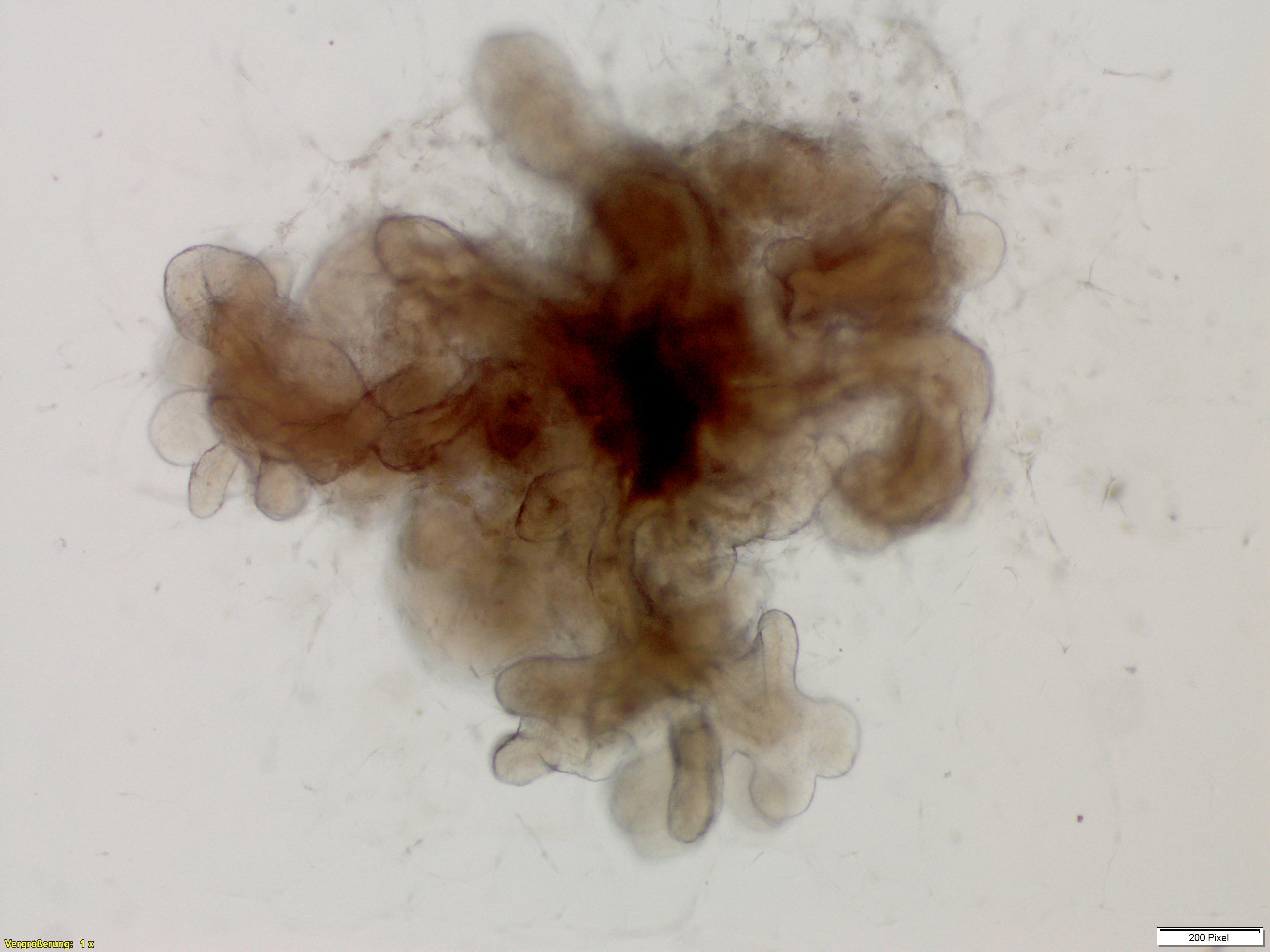
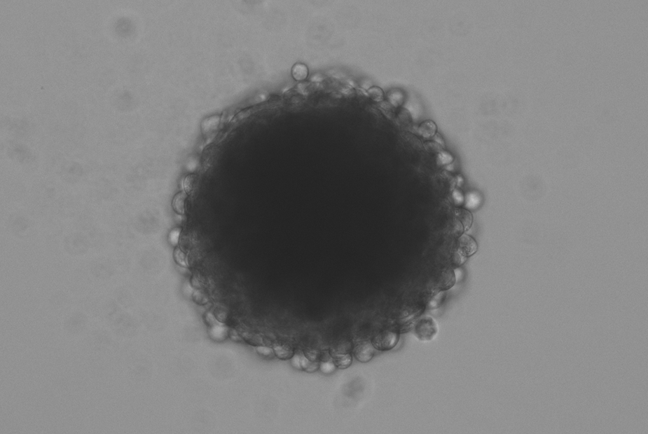
We establish organoids from stem cells and spheroids from (tumor) cell lines as multicellular 3D in-vitro microtissues. These mimic the microenvironment of cells including the specific morphological and physiological properties of complex tissues and organs.
Three-Dimensional (3D) Microtissues: Organoids and Spheroids

Organoids
An organoid is a self-organized 3D tissue that is usually derived from pluripotent, fetal or adult stem cells and mimics the essential functional, structural and biological complexity of an organ. This self-organization is specifically controlled by signaling molecules, growth factors and biological stimuli. Compared to conventional 2D cultures and animal models, organoid cultures allow for patient specificity in the model while recapitulating in-vivo tissue-like structures and functions in vitro.

Spheroids
are formed as simple, spherical 3D cell aggregates from just one cell type or a multicellular cell mixture using defined cultivation methods. Based on cancer cell lines, so-called tumoroids can also be formed, which resemble solid tumors.
Next-generation test systems for research, drug development and medicine
For decades, mammalian 2D cell cultures have been used as simple in-vitro test systems for substance testing and screening for new compounds in drug development. Organoids, however, are the “next generation” of biological tools for research, drug development and medicine. Due to their complex tissue and organ-like properties, they represent the in-vivo situation and are particularly suitable for use in translational research.
Our development: organoids and spheroids of varying complexity
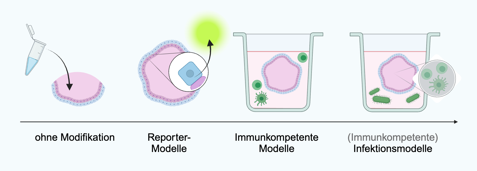
With our decades of expertise in cell culture technology and the handling of (pathogenic) microorganisms as well as working to the highest quality standards, we develop organoids and spheroids of varying complexity. Depending on the application, the 3D microtissues can also contain immune cells, microorganisms and reporter systems for detecting the activation of a cellular signaling pathway.

Our portfolio of organoids and spheroids
We provide specifically suitable 3D microtissues for different questions and applications:
- Human and animal 3D microtissues
- Reporter spheroids for non-invasive real-time detection of the activation of cellular signaling pathways
- Immunocompetent organoids
- Organoids for the investigation of microbiomes and infections with bacteria, fungi and viruses
Tabbed contents
Applications
Applications of spheroids and organoids
3D microtissues from human (stem) cells have important applications in biomedical research, as they are far superior to animal experiments in terms of their predictive value. The use of 3D microtissues as human in-vitro test systems enables
The use of 3D microtissues as human in-vitro test systems enables
- Higher throughput (using special microtiter plates for cultivating spheroids and a unique 3D incubator and reactor for cultivating organoids),
- A deeper understanding of mechanisms of action by studying specific heterogeneous cell populations, intrinsic histological structures and the communication between cells and the surrounding extracellular matrix,
- Genomic, epigenomic, pharmacological and biochemical studies and
- A deeper understanding of the development of organs, diseases and the development of cancer,
- The risk assessment of substances according to the concept of Adverse Outcome Pathways (AOP).
We provide a broad portfolio of 3D microtissues as in-vitro test systems for various applications to assess the efficacy and safety of drugs and chemicals, for
- Determination of toxicity,
- Analysis of transport processes and absorption of a substance,
- Development and preclinical testing of active ingredients and
- Investigation of the activation of cellular signaling pathways (sensitization, inflammation, ER stress)
Our services
Range of services
We offer the following services relating to our 3D microtissues, which we carry out in state-of-the-art laboratories on behalf of and in collaboration with customers:
- Conducting studies and substance screenings
- Development of innovative (reporter) spheroids
- Development of complex organoids
Analysis methods
Analysis methods
In our 3D microtissues, the effects of chemicals, active substances and products on the architecture of the tissue and physiological properties can be investigated. We offer the following analysis methods:
- Vitality measurements, for example using the MTT test (colorimetric test), alamarBlue(TM)
- Multiplex analysis for the detection and quantification of secreted proteins such as cytokines, chemokines, growth factors and antimicrobial peptides
- Histological and immunohistochemical staining of tissue thin sections
- Protein analyses using Western blot
- Analysis of specific signaling pathway activation via reporter proteins
- RNA and DNA analyses using PCR or qPCR and sequencing
 Fraunhofer Institute for Interfacial Engineering and Biotechnology IGB